Nuclear medicine is used to determine how an organ is functioning. For example, nuclear medicine can be used to study how a damaged heart is functioning or how blood flow is restricted in parts of the brain. Other organs that can be imaged using nuclear medicine include the liver, kidneys, thyroid, bones, and many others.
During a nuclear medicine study, chemicals "tagged" with radionuclides that emit gamma rays are injected into the body. The chemicals collect in the area of the body that is to be studied. Images, which may be taken immediately or days after the injection, are obtained by using a gamma camera that detects the gamma rays and produces an image map called a scintigram.
Nuclear medicine studies provide information that can help your doctor diagnose and treat diseases in their early stages. Nuclear medicine, using radioactive substances, can also be used to treat certain diseases and conditions, such as overactive thyroid glands or thyroid tumors.
Preparing for a Nuclear Medicine Study
Generally, there are no special preparations that need to be made prior to having a nuclear medicine study. However, certain types of studies may require some preparation. For example, prior to having a thyroid uptake and scan, your doctor may ask that you avoid foods and medications with iodine for one week prior to the test. Your doctor or radiologist can advise you about any special instructions you may need to follow.
Featured Locations
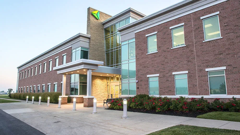
ProMedica Radiology Levis Commons
Welcome to ProMedica Radiology Levis Commons Perrysburg, Ohio. Get driving directions, hours of operation, learn about treatment and therapies and more.
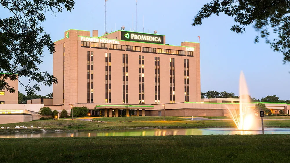
Flower Hospital
Radiology
Discover what ProMedica Flower Hospital Radiology has to offer.
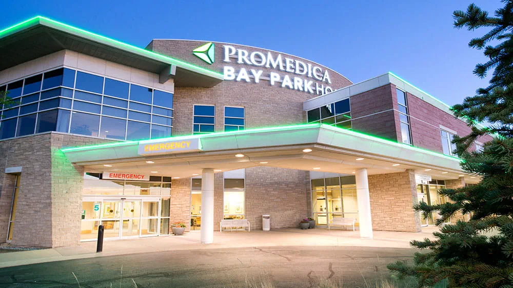
Bay Park Hospital
Radiology
Discover what ProMedica Bay Park Hospital Radiology has to offer.
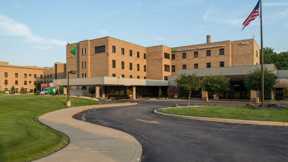
Memorial Hospital
Radiology
Discover what ProMedica Memorial Hospital Radiology has to offer.
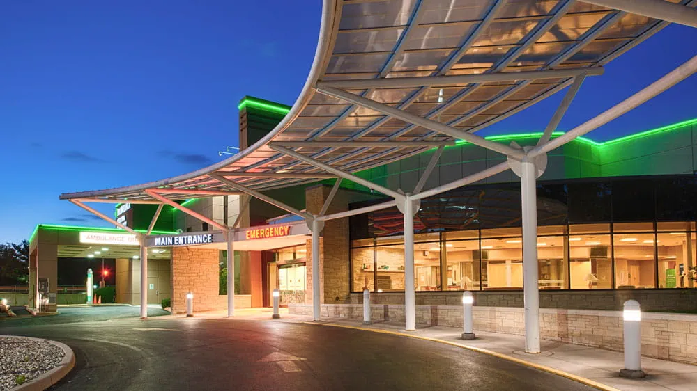
Fostoria Community Hospital
Radiology
Discover what ProMedica Fostoria Community Hospital Radiology has to offer.
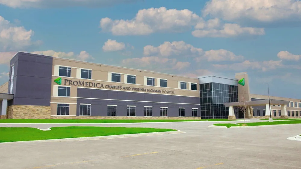
Charles and Virginia Hickman Hospital
Radiology
Discover what ProMedica Charles and Virginia Hickman Hospital Radiology has to offer.
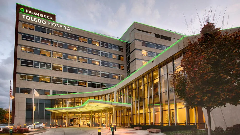
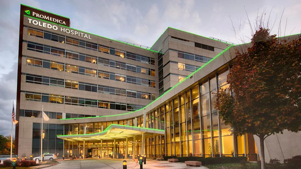
Toledo Hospital Radiology
Discover what ProMedica Toledo Hospital Radiology has to offer.

Northwest Ohio Hemophilia Center
Welcome to Northwest Ohio Hemophilia Center Toledo, Ohio. Get driving directions, hours of operation, learn about treatment and therapies and more.
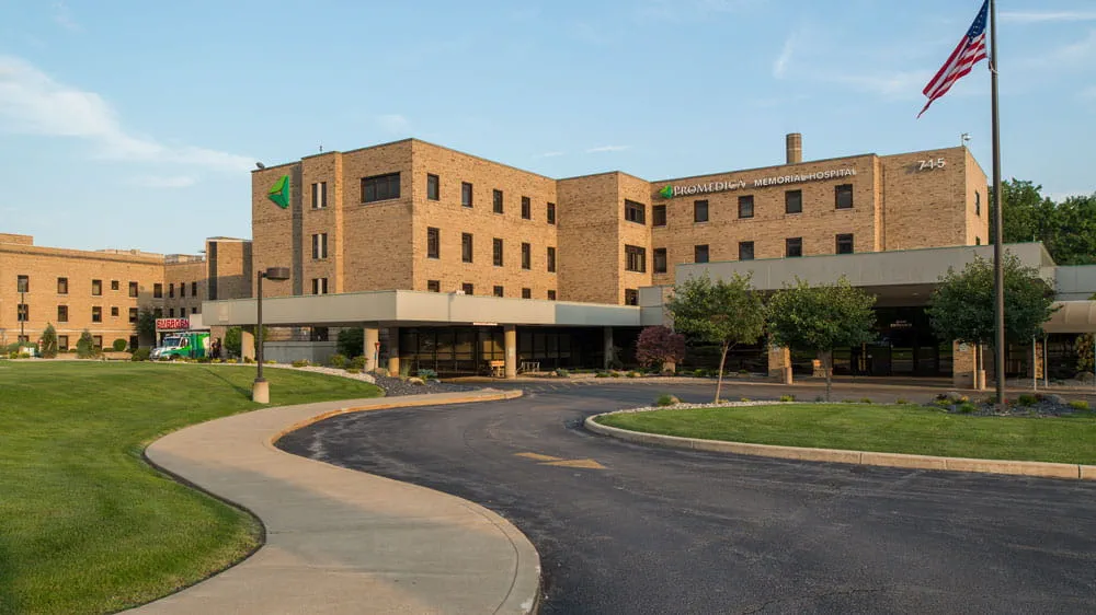
Fremont
ProMedica Memorial Hospital Women's Diagnostic Center
Advanced breast imaging, including 3D mammography, is available at ProMedica Memorial Hospital.
Oregon
Women's Imaging
Discover what ProMedica Bay Park Hospital Women's Center has to offer.
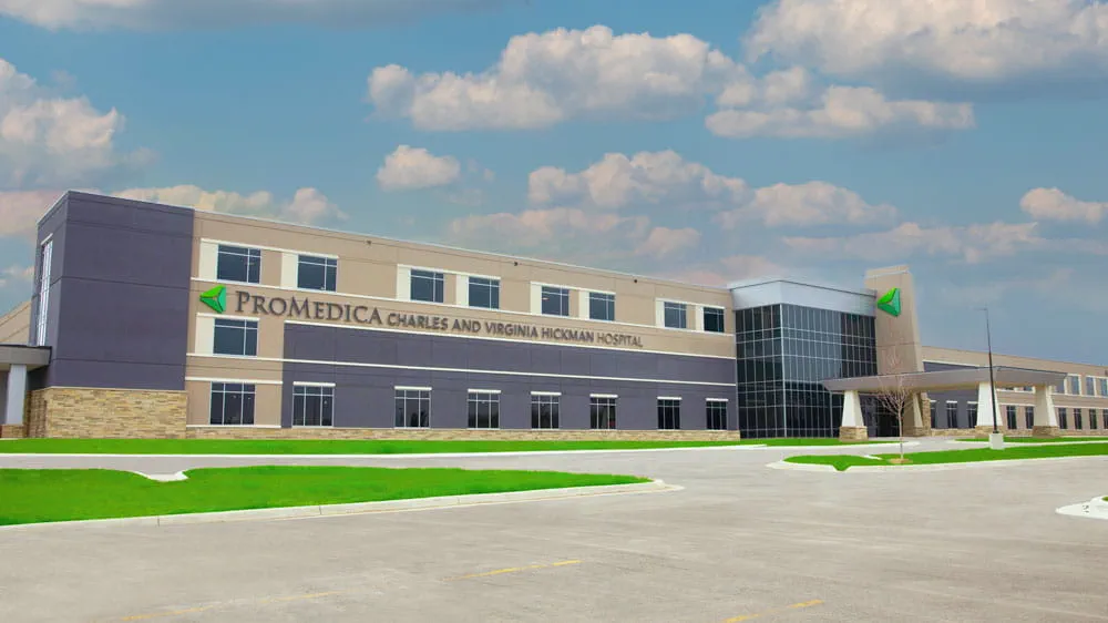
Adrian
Women's Imaging
Discover what ProMedica Charles and Virginia Hickman Hospital Women's Health Center has to offer.
Related Services & Conditions
Radiology
When you need diagnostic imaging to get a clearer picture of your health, we offer board-certified radiologists and state-of-the-art technology at 30 locations.